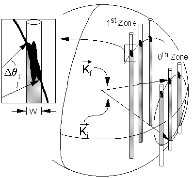
Due to the fact that only the surface is probed by RHEED the reciprocal lattice will consists of lattice rods in the direction normal to the surface probed instead of discrete spots as in electron microscopy. Figure 21 shows those lattice rods. The incoming wave vector is labeled K_i, while the diffracted beam is labeled K_f. Constructive interference occurs where K_f, defining the Ewald sphere, intersects with the reciprocal lattice rods. If a phosphorus screen is placed in front of the sample, diffraction spots will appear at positions on the screen which are given by extending the K_f's that satisfy the Bragg condition to the screen.
Ideally the reciprocal lattice rods are very thin, but due to surface irregularities those rods have a finite width as shown in the figure. Therefore the diffraction condition for a rod is given by the angle delta theta_f, which causes the ideal dots on the screen to become streaks.
If a surface is unreconstructed only integral streaks will appear on the screen, dictated by the crystal lattice. If reconstructions are present, fractional streak will appear between the integral streaks due to the fact that reconstructions usually occur as a multiple of the lattice periodicity. Rough surfaces (3-D) cause the beam to pass through facets on the sample surface, resulting in a diffraction pattern as given in a Transmission Electron Microscope (TEM).
During growth of a high quality surface oscillations in intensity of the integral streaks can be observed, which can in very simple terms be described by layer by layer growth, where a complete layer gives the brightest streaks, while a half layer somewhat disturbs the surface periodicity causing the RHEED intensity to decrease. The frequency of these oscillations correspond to the growth rate, while the amplitude of the oscillations are a function of the "smoothness" of the surface during growth.

Figure 21: RHEED Pattern (Ref. 39)
Table of Contents