11.2 Post Growth Characterization of Nitride Films - Auger Spectroscopy
Auger Electron Spectroscopy is a convenient tool for the chemical analysis
of surfaces. The Auger process is based on the fact that when an impinging
electron knocks out a core electron of an atom, the atom may decay to a
lower energy state by ejecting another electron to yield a doubly ionized
atom. The ejected electron assumes a kinetic energy very characteristic
of the atomic specie, and is equal to the difference of the singly ionized
and the doubly ionized state of the atom. Only atoms close to the surface
can eject Auger electrons without loss of kinetic energy. These characteristic
energies are superimposed on the secondary electron energy distribution.
It is therefore customary to differentiate the spectrum, so that the Auger
peaks superimposed on a rather large background can be easier detected
and identified. Approximate atomic concentrations on the surface can be
obtained by comparing the peak to peak value to a standard or by the use
of a sensitivity table. Auger is sensitive to about 0.2 atomic percent.
More information about the principles and operation of an Auger electron
spectroscopy system can be found in the Handbook published by Physical
Electronics Industries (Ref. 46), as well as spectra of nearly all elements
in the periodic table which are used to identify elements in an unknown
sample.
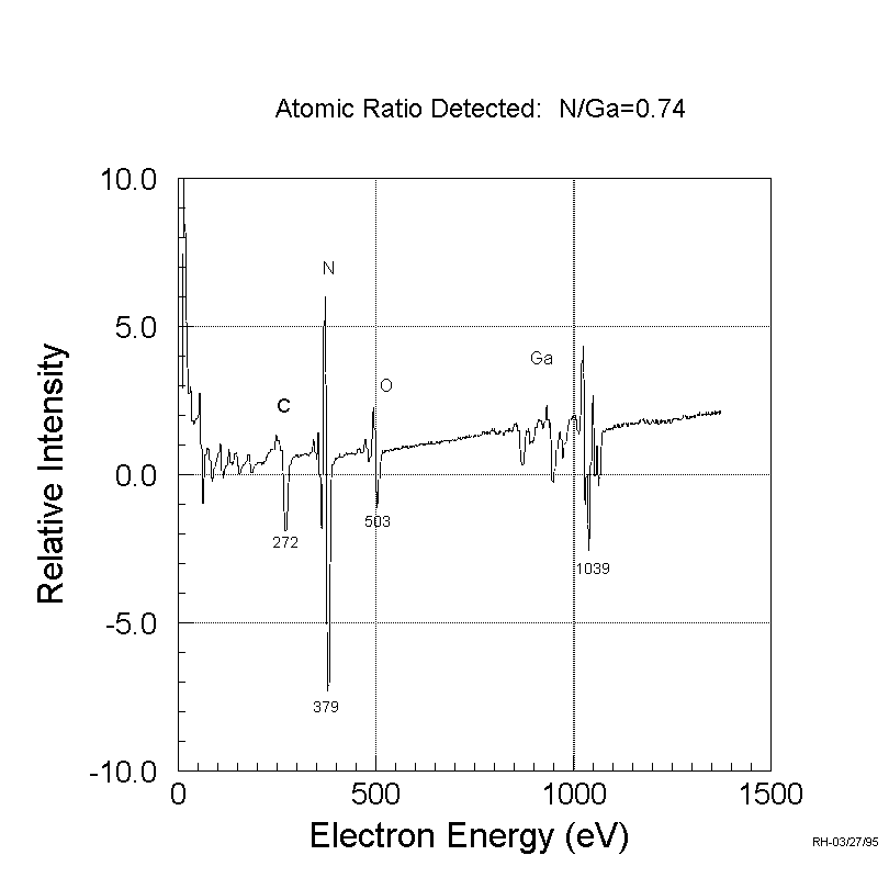
Auger Spectroscopy was routinely employed in the analysis chamber of
our vacuum system. A sample scan is shown in Figure 41, which shows besides
the Ga and N peaks also C and O. Typically C and O do not show up on the
surface, but in this case the gas handling line for the NH_3 was contaminated
and this graph was chosen to demonstrate the effect. Auger proofed to be
a valuable tool to check for contamination in our growth chamber after
growth.
The ratio of the N to Ga peak after correction for sensitivity of the
Auger system to the respective peaks was found to be about 0.75:1. This
indicates that the terminating layer is usually Ga, although the sample
was under NH_3 flux without Ga flux down to a substrate temperature of
500°C. More investigations are needed concerning this issue, which
could not be performed until recently after our Auger system was serviced.

Figure 41: Auger surface scan of GaN grown on a-plane sapphire.
Note that the atomic ratio detected does not mean that this is the
bulk concentration.
Table of Contents

